Orbital Decompression
Before and After Photo Gallery
-

-

-

-
Pic – 1
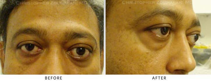
Preoperative photos a gentleman with thyroid eye disease resulting in proptosis and lower lid retraction. The left eye has more proptosis than the right eye. Note that the left lower eyelid is more retracted (lower) than the right lower eyelid.The patient underwent orbital fat decompression in the right eye. Since the left eye was more proptotic than the right eye, he also underwent bony decompression as well in addition to the orbital fat decompression (lateral and medial wall decompression).
Pic – 2
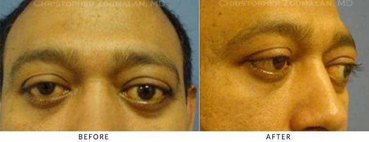
This figure above is 6 months after undergoing orbital fat decompression in the both eyes long with left lateral bony wall decompression in the left eye. He has excellent decompression with improvement in his proptosis. He still lower lid retraction which is resulting in lagophthalmos and dryness to his eyes.
Pic – 3
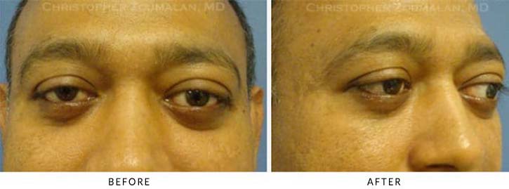
This figure is 8 months after undergoing lower lid retraction repair using hard palate grafts and canthoplasties. The hard palate graft is a spacer that allows to raise the lower lid by acting as a support for the lower lid. They are taken from the patient’s own roof of their mouth. The hard palate is an excellent source of a posterior spacer graft. Other alternatives include Alloderm and cartilage grafts. I prefer using either hard palate or Alloderm grafts in my patients. The canthoplasties were down to resuspend the lids laterally to allow for a better improvement in retraction in addition to the hard palate grafts.
-
Patient Seeing Side
-
Disclaimer:
*Individual results may vary
All before and after pictures displayed are real patients who have consented to having their pictures published on our site. Individual results will vary with each patient and Dr. Christopher Zoumalan does not guarantee any outcomes of procedures shown. All pictures are meant for reference and illustrative purposes only.


